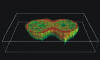
Examples of 3-D reconstructions of
intracellular
fluorescent distributions
Examples of spatial distribution of the cytoskeletal proteins obtained by confocal laser scanning microscopy and visualized by the software package amira.
| I M A G E S | |
|
|
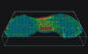
Distribution of the motor protein myosin II in the final stage of cytokinesis in the protist Dictyostelium discoideum (immunofluorescence) |
 |
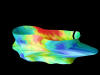
Distribution of the protein cortexillin during the cell division of a wild-type Dictyostelium cell (sagittal section) |
 |
Distribution of the protein cortexillin during the cell division of a Dictyostelium cell that lacks myosin II (sagittal section) |
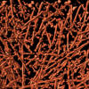 |
Actin filaments in the cortical cytoskeleton of a Dictyostelium discoideum cell (cryoelectron tomography) |
|
|
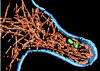
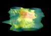
Granular distribution of a protein Nm23/NDPK subunit in a tumor cell (fusion with green fluorescent protein GFP) |
|
|
Structure of a nascent filopodium: actin filaments - RED, cell plasma membrane - BLUE, cell organelles - GREEN (cryoelectron tomography) |
|
|
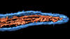
Structure of a mature filopodium: actin filaments - RED, cell plasma membrane - BLUE (cryoelectron tomography) |
| A N I M A T I O N S | |
 |
Animated spatial distribution of a protein Nm23/NDPK subunit in a tumor cell [0.65 Mb] |
|
|
Virtual stroll through the cytoplasm: actin filaments - RED, intracellular membranes - DARK BLUE, ribosomes - LIGHT BLUE (cryoelectron tomography) [5 Mb] |
 |
3-D visualization of the
cortexillin enrichment in the cleavage furrow of a dividing cell [0.8 Mb] |
 |
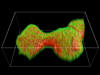
The cortexillin enrichment time course displayed as an 3-D envelope (the time axis runs upwards) [2.9 Mb] |